

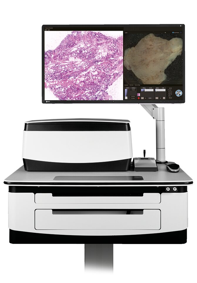
VivaScope 2500
The VivaScope 2500 uses two lasers with wavelengths of 488 nm (blue) and 638 nm (red). A fluorescent dye that is applied to the tissue prior to the VivaScope imaging process is excited by the blue laser, thus highlighting cell structures (e.g. nuclei). Additionally, the red laser is used to generate a reflectance signal, showing structural information of the sample. Both reflectance and fluorescence signals are acquired simultaneously and correlated in real-time.
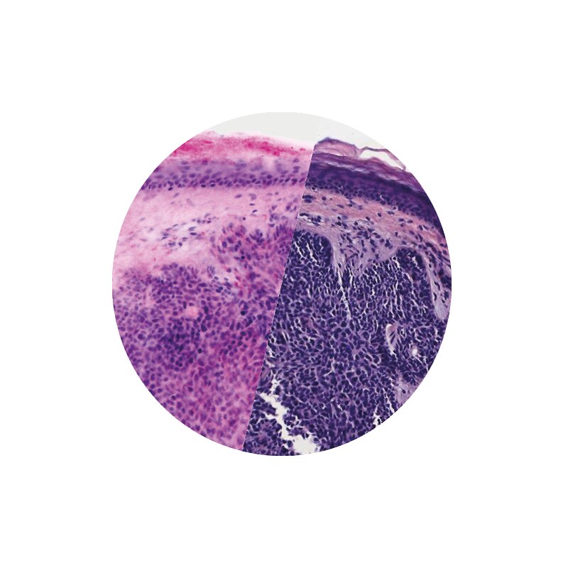
A built-in algorithm translates the signals into H&E-like pseudocolored images. The resulting images contain similar information to conventional histology and can be examined at any desired magnification, ranging from displaying the whole sample up to a 550-fold magnification.
Reviews
Our advantages

Only high quality
products

Authorized
representative and
own production

Individual and
approach

Qualified
consultation

Large warehouse and
fast delivery
in Ukraine


